
The pressure distribution in the auditory canal in a progressive sound field. The effect of tympanic muscles on sound transmission. Tympanic-membrane vibrations in human cadaver ears studied by time-averaged holography. Re-examination of the role of the human acoustic stapedius reflex. Effect of mastoid cavity modification on middle ear sound transmission. Physiology of the eustachian tube and middle ear pressure regulation.
#Auditory ossicles function update#
Update on anatomy, development, and function. Are two muscles needed for the normal functioning of the mammalian middle ear?. Measurement of the ossicular vibration ratio in human temporal bones by use of a video measuring system. Effect of tensor veli palatini muscle paralysis on eustachian tube mechanics. A quantitative study of the effect of the acoustic stapedius reflex on sound transmission through the middle ear of man. The Role of the Clinically Obtained Acoustic Reflex as a Research Tool for Subclinical Hearing Pathologies. 45(7):417-27.Ĭauson A, Munro KJ, Plack CJ, Prendergast G.

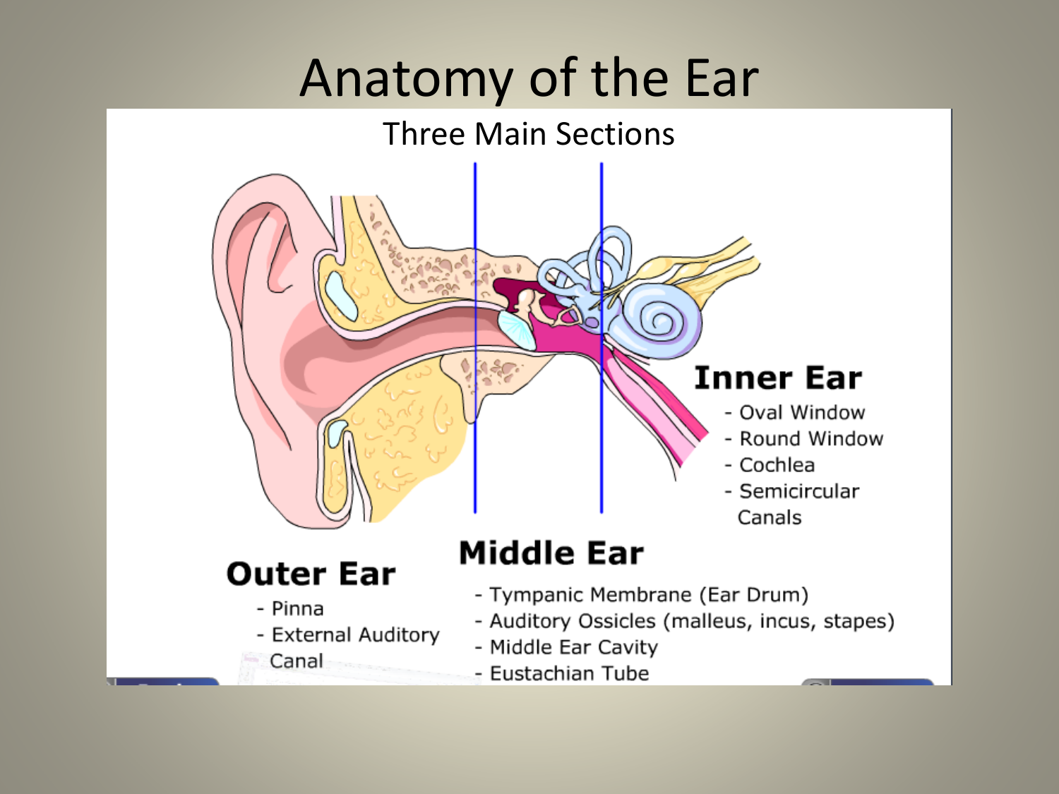
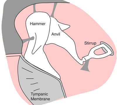
Choice of probe tone and classification of trace patterns in tympanometry undertaken in early infancy. Reversed ipsilateral acoustic reflex pattern. Bilateral tinnitus due to middle-ear myoclonus. Audiological long-term results following stapedotomy with stapedial tendon preservation. Combined effect of fluid and pressure on middle ear function. Successful treatment of autophonia with botulinum toxin: case report. Finite element analysis of active Eustachian tube function. A human temporal bone study of stapes footplate movement. Analysis of middle ear mechanics and application to diseased and reconstructed ears. Histological and Histopathological Study of Incus. Morphology and function of Neandertal and modern human ear ossicles. Stoessel A, David R, Gunz P, Schmidt T, Spoor F, Hublin JJ. Effects of changes in dynamic characteristics of the middle ear on transient-evoked otoacoustic emissions. Spiric S, Spiric P, Vranjes D, Aleksic A. Conditions for Highly Efficient and Reproducible Round-Window Stimulation in Humans. Schraven SP, Hirt B, Goll E, Heyd A, Gummer AW, Zenner HP, et al. Viscoelastic properties of human tympanic membrane. Sound pressure generated in an external-ear replica and real human ears by a nearby point source. Theoretical and applied external ear acoustics. Binaural weighting of pinna cues in human sound localization. Sound pressure gain produced by the human middle ear. Any dysfunction or disease of these components results in a conductive hearing loss, and clinically, an individual’s inability to properly hear sound. Proper impedance matching requires the normal anatomy and functioning of an external ear and a middle ear with an intact tympanic membrane, a normal ossicular chain, and a well-ventilated tympanic cavity. If no middle ear were present, only 0.1% of the acoustic wave energy traveling through air would enter the fluid of the cochlea and 99.9% would be reflected.įor physiologic hearing to occur, a precochlear amplification system must be present to address the impedance mismatch that exists between air and water. When a sound wave is transferred from a low-impedance medium (eg, air) to one of high impedance (eg, water), a considerable amount of its energy is reflected and fails to enter the liquid. Interestingly, they are the only bones in the body that do not grow after birth and are also the smallest bones in the body ( variant tiny sesamoids aside).The primary function of the middle ear is to offset the decrease in acoustic energy that would occur if the low impedance ear canal air directly contacted the high-impedance cochlear fluid. The stapes also articulates with the oval window via the stapediovestibular joint, which is a syndesmosis 3 this joint transmits the ossicular vibrations to the endolymph in the vestibule. They are located in the middle ear cavity and articulate with each other via two tiny synovial joints. Their role is to mechanically amplify the vibrations of the tympanic membrane and transmit them to the cochlea where they can be interpreted as sound. There are three tiny articulating bones in the middle ear known as ossicles (from lateral to medial):


 0 kommentar(er)
